Summary
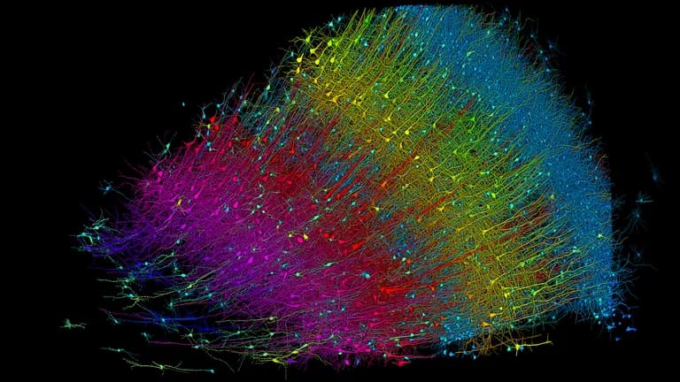
Researchers charted a cubic millimeter volume of the human brain at nanoscale resolution. They traced every neuron, synapse, blood vessel, and supporting cell within the fragment. Though it represents just one-millionth of the total brain volume, it is the most detailed map of a piece of human brain ever created.
Researchers built a 3D image of nearly every neuron and its connections within a small piece of human brain tissue. The segment’s neuron density is 16,000 neurons per cubic millimeter. The reconstructed segment is 1.4 petabytes in digital size, or 1,400 terabytes.
An intriguing finding in the reconstruction was the existence of clusters of cells that tended to occur in mirror-image orientation to one another. The scientists further found a new type of unexplained structure that they’ve named an “axon whorl,” wherein long axon cables appear to tangle around themselves.
The team has ambitions to construct multiple partial connectomes representing additional human brain samples. They are also working on zebrafish connectome, and are planning to tackle increasingly large segments of the mouse brain. Mammalian brains share many similarities, so a complete mouse connectome could offer new insights into our own brain.
